

Meiosis
Meiosis
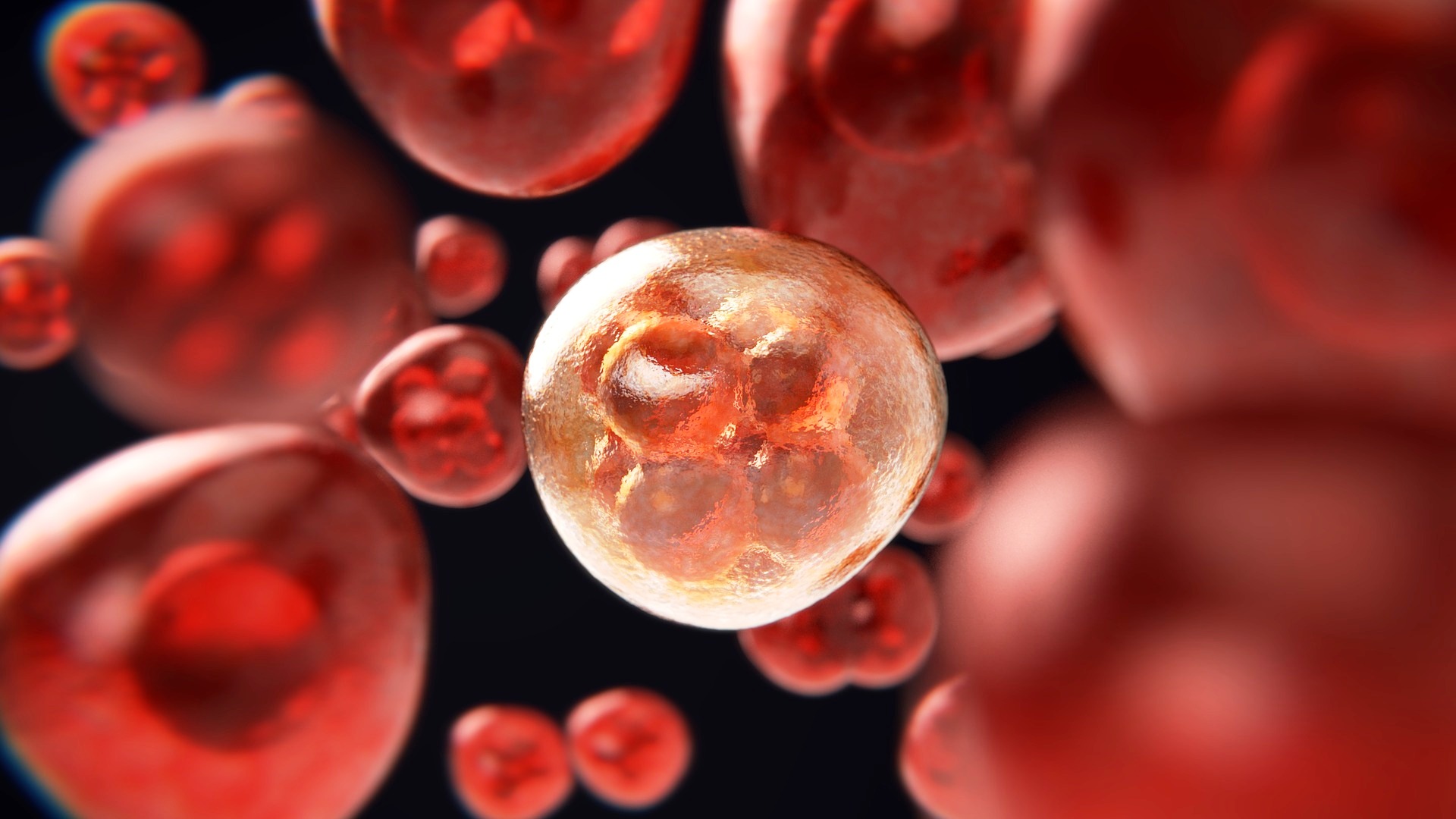
With the term meiosis, in biology, we mean the process of cell division that exclusively concerns the reproductive cells, where 4 daughter cells are formed from a mother cell, all different from each other containing half of the parent’s genetic heritage.
An example of meiosis occurs in sexual reproduction in Angiosperms, where the fusion between the male gamete (spermatic nucleus of the microgametophyte) and the female one (egg cell of the macrogametophyte) takes place; in this event it is necessary that these two cells have half the genetic heritage of the diploid somatic cell (2n), i.e. they are haploid (n), to originate from their fusion (gamia) the zygote with genetic heritage (2n) consisting of the halved kits and remix of both parents.
With meiosis, in this way, genetic variability is guaranteed and the doubling of the genetic patrimony with each sexual reproduction is avoided.
In animals, meiosis occurs in the gonads, during the maturation of germ cells (gametogenesis): gametes are haploid, all other cells diploid (gametic or terminal meiosis).
In most plants, it occurs in an intermediate stage of the cycle (sporic or intermediate meiosis), so that there is an alternation of generations between a diploid stage (sporophyte or diplophyte) and a haploid stage (gametophyte or aplophyte).
In some Protozoa and in many lower plants (Chlorophytes, Ficomycetes, Ascomycetes, Basidiomycetes), meiosis is zygotic or initial, that is, it occurs immediately after fertilization, so that only the zygote is diploid, and all the other stages are represented by cells haploid. A nucleus with a diploid chromosomal set contains two copies of each chromosome, which come one from the father and the other from the mother except for the two sex chromosomes of the heterogametic sex (for example, the X and Y chromosomes of man); the chromosomes of a pair are called homologous.
Genetic variability –
Meiosis is the process underlying genetic variability; new genetic combinations can be formed through two fundamental processes: a) homologous chromosomes are distributed in different combinations among the haploid products of meiosis by a process called independent assortment of chromosomes. For example, from a diploid cell containing three pairs of homologous chromosomes, 1M and 1P, 2M and 2P, 3M and 3P (where M indicates the maternal chromosomes and P the paternal chromosomes), haploid gametes with 8 possible different chromosomal combinations will be formed.
Homologous chromosomes take part in the genetic exchange, during meiotic prophase, by crossing-over. A typical haploid gamete will therefore contain chromosomes derived from the mother, chromosomes derived from the father, and some recombinant chromosomes that contain information derived from both the paternal and maternal chromosomes.
This cell, with its specific and unique genetic combination, will fuse with a second haploid cell, also with its specific genetic combination, to produce the 2n diploid zygote which will have a different genetic composition from that of the two diploid individuals from which derives.
Stages of Meiosis –
The meiosis process is preceded by a phase in which the synthesis of DNA (S) takes place; during meiosis the number of chromosomes of a 2n diploid cell is reduced to half by two successive nuclear divisions: in the first division the homologous chromosomes, made up of two chromatids, separate into the two daughter cells, which receive only one element of the pair. In the second meiotic division the two chromatids of each chromosome are separated to form the 4 haploid gametes. Each of the two divisions is characterized by 4 successive phases, called: prophase, metaphase, anaphase and telophase.
Prophase –
The prophase of the first meiotic division (prophase I) is divided, in turn, into various stages. In the first (leptotene) the chromosomes are identified under an optical microscope and appear as thin filaments (chromomemes) in which intensely colorable granules (chromomers) are distinguishable. Each chromosome adheres with both its ends to the nuclear envelope by means of an attachment plate. In the second stage (zygotene) the homologous chromosomes pair up by approaching along their entire length with a ‘zip closure’ process that ensures the pairing of each allele with its homologue on the opposite chromosome. The synapse is preceded by the formation of a filiform protein axis along each of the homologues; during pairing, the protein axes adhere to form a ladder-shaped structure, called the synaptine complex. Each pair of chromosomes is called bivalent but, in this stage, 4 chromatids are visible for each bivalent which therefore can be called tetrad. The so-called pachytene stage follows, in which the chromosomes shorten and thicken, always remaining paired. In this phase the crossing-overs determine the exchange between chromatids belonging to the two different homologous chromosomes. In the next stage (diplotene), the synaptine complex breaks down allowing the separation of the two homologous chromosomes; however they remain united by one or more chiasms, the sites where the crossing-over took place.
In oocytes, the diplotene stage can last for months or years because it is precisely at this stage that the chromosomes despiralize and begin the synthesis of RNA, which will serve to provide the egg’s reserve material. Diakinesis is the transition stage towards metaphase when RNA synthesis stops and the densified chromosomes detach from the nuclear envelope. Each bivalent shows the 4 chromatids of which it is composed: the child chromatids of each chromosome are joined together at the centromere level, while the chromatids of the homologous chromosomes that have undergone crossing-over are linked by chiasms.
At the end of the long and complex prophase I, meiosis continues with two subsequent nuclear divisions which, overall, occupy only 10% of the total time required for the entire process.
Metaphase, anaphase, telophase –
In metaphase I, the bivalents are arranged on the spindle (on the equatorial plate of the cell) and the centromeres of the two elements of each bivalent are oriented towards the opposite poles of it.
In anaphase I, the centromeres move away towards the poles of the spindle and the chiasms move towards the free end of the chromatids, a phenomenon called terminalization of the chiasms.
In telophase I, 2 nuclei are formed with a haploid number of chromosomes.
Each chromosome consists of the two chromatids joined by the centromere.
Interkinesis, with the despiralization of chromosomes and the formation of two cells, is a stage that does not always occur and is in any case short-lived.
Metaphase II follows in which the n elements of each nucleus, with only the centromere still undivided, are arranged on the equatorial plate. The centromere divides and thus passes to anaphase II, in which the centromeres move to opposite poles of the spindle and the chromatids of each chromosome divide.
At telophase II there are, as descendants of the original cell, 4 cells with haploid number (n) of chromosomes. In female gametogenesis the 4 cells produced are the mature egg and the 3 polar blood cells, in male gametogenesis they are the 4 spermatids.
Anomalies of Meiosis –
Like all biochemical processes, meiosis can also undergo factors that modify or alter its course.
Alterations can thus occur, some of which can even have serious consequences.
The most serious alteration during the meiosis process is that of the so-called non-disjunction, that is, the failure to separate a pair of chromosomes (or a pair of chromatids).
This alteration was detected for the first time in the X chromosomes of Drosophila and is linked in humans to numerous syndromes: Down syndrome is associated with the nondisjunction of the chromosomes of the pair 21; Klinefelter’s and Turner’s syndrome of the pair of the two X chromosomes; Pätau syndrome (trisomy 13) of couple 13 and Edwards syndrome (trisomy 18) of couple 18.